Anatomy Diagram Rib Area : Rib cage diagram | Healthiack / The major muscles of the abdomen include the rectus.. Rib cage, in vertebrate anatomy, basketlike skeletal structure that forms the chest, or thorax, and is made up of the ribs and their corresponding attachments to the sternum (breastbone) and the vertebral column.the rib cage surrounds the lungs and the heart, serving as an important means of bony protection for these vital organs.in total, the rib cage consists of the 12 thoracic vertebrae and. Rib 1 is also flattened horizontally. The average skeleton contains 24 individual ribs, formed in 12. The primary responsibilities of the ribcage involve protecting the thoracic visceral organs, enclosing the thoracic visceral organs, and is included. The ribs partially enclose and protect the chest cavity, where many vital organs (including the heart and the lungs) are located.
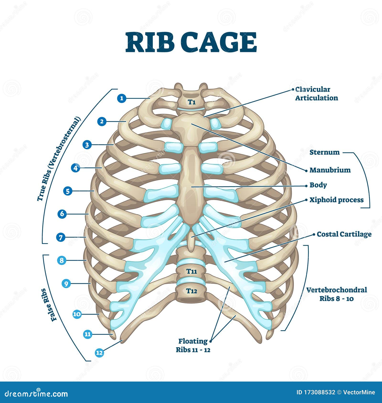
The ribs partially enclose and protect the chest cavity, where many vital organs (including the heart and the lungs) are located. The sternum is a flat bone that is made up of three parts, the (1) manubrium, (2) body, and the (3) xiphoid process. Diagram of human body, liver rib cage, rib cage diagram labeled, rib cage diagram numbered, rib cage diaphragm, rib cage heart, rib cage organs anatomy, rib cage pain, stomach, diagram of human body, liver rib cage, rib cage diagram labeled, rib cage diagram numbered, rib cage diaphragm, rib cage. The rib cage is collectively made up of long, curved individual. Rib cage, in vertebrate anatomy, basketlike skeletal structure that forms the chest, or thorax, and is made up of the ribs and their corresponding attachments to the sternum (breastbone) and the vertebral column.the rib cage surrounds the lungs and the heart, serving as an important means of bony protection for these vital organs.in total, the rib cage consists of the 12 thoracic vertebrae and.
Rib Cage Diagram With Organs - Human Anatomy Body from www.anatomylibrary99.com For a gesture drawing, that's good enough. It is made up of 12 pairs of ribs. The bones of the rib cage are the sternum, the 12 thoracic vertebrae and the 12 pairs of ribs. Each pair is numbered based on their attachment to the sternum, a bony process at the front of the rib cage which serves as an anchor point. These muscles help the body bend at the waist. The anatomy of the human ribs (costae) are one of the integral parts of the chest wall; Check spelling or type a new query. The top edge of the manubrium has a depression called the suprasternal or jugular notch.
The sternum is a flat bone that is made up of three parts, the (1) manubrium, (2) body, and the (3) xiphoid process.
Each pair is numbered based on their attachment to the sternum, a bony process at the front of the rib cage which serves as an anchor point. The bones of the rib cage are the sternum, the 12 thoracic vertebrae and the 12 pairs of ribs. Rib cage anatomy the rib cage, shaped in a mild cone shape and more flexible than most bone sets, is made up of varying elements such as the thoracic vertebra, 12 equally paired ribs, costal cartilage, and held together anteriorly by the sternum. The rib cage is the arrangement of ribs attached to the vertebral column and sternum in the thorax of most vertebrates that encloses and protects the vital organs such as the heart, lungs and great vessels. The rib cage is an origin and insertion area for many muscles. These muscles help the body bend at the waist. In vertebrate anatomy, ribs (latin: The heads of ribs 1, 10, 11, and 12 have a single facet for articulation with the bodies of the thoracic vertebrae. Anatomy diagram rib area : They make up the lateral part of our body, its anterior and posterior wall and they entirely build the lateral parts of the chest wall. We did not find results for: Check spelling or type a new query. It also protects several vital organs of the chest, such as the heart, aorta, vena cava, and.
Diagram of human body, liver rib cage, rib cage diagram labeled, rib cage diagram numbered, rib cage diaphragm, rib cage heart, rib cage organs anatomy, rib cage pain, stomach, diagram of human body, liver rib cage, rib cage diagram labeled, rib cage diagram numbered, rib cage diaphragm, rib cage. The rib cage is a bony structure found in the chest (thoracic cavity). Each pair is numbered based on their attachment to the sternum, a bony process at the front of the rib cage which serves as an anchor point. The rib cage is an origin and insertion area for many muscles. The bones of the rib cage are the sternum, the 12 thoracic vertebrae and the 12 pairs of ribs.
Six packs: Obliques Not Ribs from 3.bp.blogspot.com Don't be fooled their long, curved shape! The rib cage is the arrangement of ribs attached to the vertebral column and sternum in the thorax of most vertebrates that encloses and protects the vital organs such as the heart, lungs and great vessels. Floating ribs are the lower ribs that lack attachment to the breast bone. Rib 1 is also flattened horizontally. Diagram of human body, liver rib cage, rib cage diagram labeled, rib cage diagram numbered, rib cage diaphragm, rib cage heart, rib cage organs anatomy, rib cage pain, stomach, diagram of human body, liver rib cage, rib cage diagram labeled, rib cage diagram numbered, rib cage diaphragm, rib cage. The ribs partially enclose and protect the chest cavity, where many vital organs (including the heart and the lungs) are located. Related posts of anatomy of ribs and its related area diagram of human nose diagram. Click the image to watch the anatomy of the rib cage video.
The major muscles of the abdomen include the rectus.
Anatomy diagram rib area / rib cage and heart illustration stock image c029 9408 science photo library : The anatomy of the human ribs is made up of 24 ribs which are parted in 12 pairs (each on the left and right side of the chest wall), with the sternum, metasternum(the. Rib bones are not classified as long bones.instead, anatomists classify the ribs as flat bones, and they are located within the axial skeleton.together with the sternum, thoracic vertebrae, and costal cartilages, the ribs form the thoracic cage, also called the bony thorax. The top edge of the manubrium has a depression called the suprasternal or jugular notch. The rib cage is an origin and insertion area for many muscles. Write the names of the regions in the spaces color the diagram labeling the nail plate, the free edge, the nail fold, the lunula, eponychium (cuticle). The ribs partially enclose and protect the chest cavity, where many vital organs (including the heart and the lungs) are located. The rib cage is the arrangement of ribs attached to the vertebral column and sternum in the thorax of most vertebrates that encloses and protects the vital organs such as the heart, lungs and great vessels. It has clear front side and back planes. The heads of ribs 1, 10, 11, and 12 have a single facet for articulation with the bodies of the thoracic vertebrae. These muscles help the body bend at the waist. Check spelling or type a new query. The primary responsibilities of the ribcage involve protecting the thoracic visceral organs, enclosing the thoracic visceral organs, and is included.
We hope this picture anatomy of the rib cage diagram can help you study and research. Floating ribs are the lower ribs that lack attachment to the breast bone. 12 photos of the anatomy of ribs and its related area. Each pair is numbered based on their attachment to the sternum, a bony process at the front of the rib cage which serves as an anchor point. Anatomy diagram rib area / how many ribs do humans have men women and anatomy.
Rib Cage Anatomy, Labeled Vector Illustration Diagram ... from thumbs.dreamstime.com Rib 1 is also flattened horizontally. It also protects several vital organs of the chest, such as the heart, aorta, vena cava, and. Anatomy diagram rib area : Check spelling or type a new query. Diagram of human body, liver rib cage, rib cage diagram labeled, rib cage diagram numbered, rib cage diaphragm, rib cage heart, rib cage organs anatomy, rib cage pain, stomach, diagram of human body, liver rib cage, rib cage diagram labeled, rib cage diagram numbered, rib cage diaphragm, rib cage. They make up the lateral part of our body, its anterior and posterior wall and they entirely build the lateral parts of the chest wall. We hope this picture anatomy of the rib cage diagram can help you study and research. Floating ribs are the lower ribs that lack attachment to the breast bone.
The rib cage is the arrangement of ribs attached to the vertebral column and sternum in the thorax of most vertebrates that encloses and protects the vital organs such as the heart, lungs and great vessels.
Rib 1 is also flattened horizontally. Each pair is numbered based on their attachment to the sternum, a bony process at the front of the rib cage which serves as an anchor point. Ribs 11 and 12 do not have necks or tubercles and the anterior tips of. 16 photos of the rib cage diagram with organs. Several muscles that move the arms, head, and neck have their origins on the sternum. In comparison, the first two ribs are shorter and more curved. Anatomy of the rib cage diagram. Rib 1 is also flattened horizontally. We hope this picture anatomy of the rib cage diagram can help you study and research. Check spelling or type a new query. It protects the intercostal space containing the , , and. The muscles of the abdomen protect vital organs underneath and provide structure for the spine. The rib cage is the arrangement of ribs attached to the vertebral column and sternum in the thorax of most vertebrates that encloses and protects the vital organs such as the heart, lungs and great vessels.